Complete Hip Replacement
Complete Hip Replacement (CHR) is an excellent cure option for people in their advanced stage of the degenerative hip disease. A CHR for these patients can bring a considerable decrease in pain with most of the patients regaining all the normal range of motions and their physical capability after the completion of the recuperative process.
Complete Hip Joint Replacement is only done when patients undergoing non-operative treatments fail to achieve respite from arthritis symptoms. Under a CHR, the procedure actually involves surgically removing the arthritic parts of the joint (cartilage & bone), replacing the "ball & socket" part of the joint with artificial components prepared from metal alloys, and inserting high-performance bearing surface between the metal parts.
After the operative procedure, a patient usually gets to spend some days in the hospital 5 to 10 days at the max in most cases. Post surgery, most of the patients have to use a walker for an initial period of 4-6 weeks, later on, they would resort to walking with the help of a walking stick for another 4-6 weeks before they can start their normal movement without any help or support.
Anatomy of a Hip Joint…
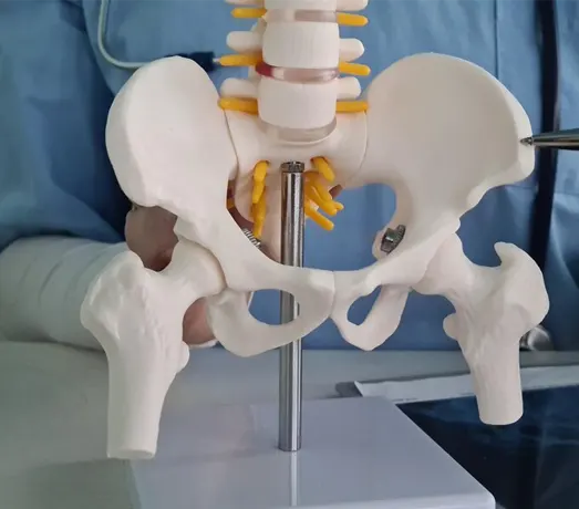
Hip joint is typically a real ball-and-socket joint of the body. The hip socket which is called the acetabulum makes a deep cup like shape to surround the ball of the upper thigh bone, known as femoral head. The thick muscles of buttocks in the back and thick muscles of thighs in the front enclose the hip.
The exterior of the femoral head and the interior of the acetabulum are enclosed with articular cartilage. This material is about a quarter inch deep in nearly all big joints. Articular cartilage is a strong, smooth substance that lets the exteriors to rub alongside one another without inflicting any injury.

What do we need an artificial Joint?
What makes an artificial Hip?
Primarily, there are 2 types of artificial hip replacements in practice.
- Cemented prosthesis
- Uncemented prosthesis
Under a cemented prosthesis procedure, the joint is put in place with the help of epoxy cement that joins the metal to the bone, while an uncemented prosthesis has an excellent net of holes on the exterior that allows bone to grow into the net and join the prosthesis to the bone. The assessment about whether a cemented or an uncemented artificial hip is to be used is normally done by the surgeon depending upon the age factor of the patient, and surgeon’s own past experience in doing such operative procedures.
